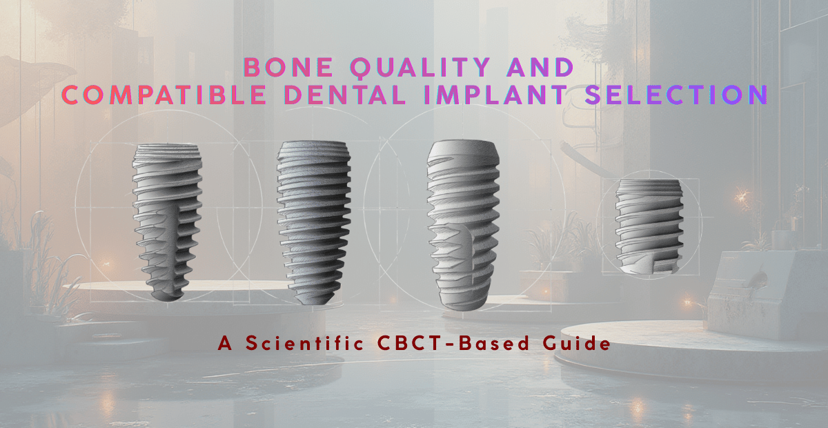
Bone Quality & Dental Implant Selection | CBCT-Based Scientific Guide
Bone Quality, Bone Quantity, and Compatible Dental Implant Selection
A Research-Based Clinical Decision Framework
Bone quality and bone quantity form the cornerstone of every successful dental-implant treatment plan. Modern implantology no longer depends solely on tactile sensation or two-dimensional radiographs. Instead, clinicians integrate classical bone classifications, three-dimensional CBCT imaging, and quantitative density analysis to select the optimal implant macro-design, surface characteristics, and surgical protocol for each patient.
This evidence-based approach improves primary stability, osseointegration, and long-term implant survival, particularly in compromised bone situations.
1. Classical Bone-Type Classifications
|
Classification |
Description of Bone Pattern |
Clinical Implant Considerations |
|
Lekholm & Zarb (Types I–IV) |
Type I: dense cortical bone; Type II: thick cortical + dense trabecular; Type III: thin cortical + porous trabecular; Type IV: very thin cortical + low-density trabecular |
Type I–II allow standard drilling and conventional implants; Type III benefits from under-preparation or tapered implants; Type IV carries the highest failure risk and often requires augmentation, wider implants, or enhanced surfaces |
|
Misch & Judy (1985) |
Class I–IV based on ridge width and height |
Guides ridge-splitting, GBR, sinus augmentation, or block grafting decisions |
|
Periobasics (Class I–VI) |
Ranges from dentate ridge (Class I) to severely resorbed, depressed ridge (Class VI) |
Serves as a surgical decision tree: Class III → standard protocol; Class IV–V → ridge expansion/GBR; Class VI → staged reconstruction |
These systems remain clinically relevant for initial assessment and surgical staging, but they lack quantitative density evaluation.
2. Quantitative CBCT-Based Bone Classification(A1–C3)
Recent CBCT-based research introduced a dual-axis classification separating cortical and cancellous components, generating nine bone types (A1–C3).
Cortical Bone Grades
- A: Thick, dense cortical shell (≥ 1200 HU) – excellent primary stability
- B: Moderate cortical thickness (≈ 800–1200 HU)
- C: Thin or absent cortical layer (< 800 HU)
Cancellous Bone Grades
- 1: High trabecular density (≈ 600–800 HU)
- 2: Moderate density (≈ 400–600 HU)
- 3: Low density (< 400 HU)
Population distribution in large CBCT samples shows B2 as the most common type, while extreme combinations such as A3 or C1 highlight why traditional systems may oversimplify bone biology.
This quantitative model allows clinicians to precisely tailor implant diameter, length, taper, thread design, and surface treatment based on the cortical-cancellous relationship rather than relying on subjective judgment alone.
3. Imaging and Analytical Tools in Implant Planning
|
Tool |
What It Provides |
Clinical Relevance |
|
Cone-Beam CT (CBCT) |
3-D bone volume, cortical thickness, trabecular density, HU values |
Enables quantitative classification and accurate implant planning |
|
Digital periapical radiographs |
Bone height and width (2-D) |
Screening tool; limited for density evaluation |
|
ISQ & Insertion Torque (IPT) |
Real-time primary stability measurements |
Strongly correlates with moderate-density bone (D2/B2) |
|
Micro-CT & histomorphometry (research) |
Trabecular orientation, collagen and apatite alignment |
Explains biomechanical adaptation and load transfer patterns |
4. Implant Selection According to Bone Type
|
Bone Type |
Recommended Implant Design |
Surgical Adjuncts |
|
Dense cortical bone (A, Type I–II) |
Standard diameter (3.5–4.5 mm), 8–12 mm length, parallel or mildly tapered, SLA or Active surface |
Conventional drilling protocol |
|
Moderate density bone (B2, Type III) |
Tapered or wider implants (4.5–5.0 mm), longer length, moderately rough or anodized surfaces |
Under-drilling, bone condensation, minor GBR if width < 6 mm |
|
Low-density bone (C3, Type IV) |
Aggressive thread design, longer implants (≥ 13 mm), nano-rough or calcium-phosphate surfaces |
Staged GBR, sinus lift, ridge expansion, delayed loading |
|
Severely compromised bone |
Wide-diameter (≥ 5 mm) implants with deep threads and platform switching |
GBR with membrane, particulate grafts, PRF |
|
Immediate post-extraction sockets |
Conical implants engaging apical bone |
Socket preservation grafting and non-occluding provisionalization |
5. Clinical Outcomes Linked to Bone Quality
- Type IV bone consistently shows lower implant survival, reinforcing the need for augmentation and surface-enhanced implants
- Moderate-density bone (D2/B2) demonstrates the highest ISQ and insertion torque values
- Advanced surface technologies significantly improve osseointegration in low-density bone by enhancing osteoconduction and early bone contact
6. Practical Clinical Workflow
- Clinical ridge assessment (Periobasics / Misch classification)
- CBCT evaluation with HU measurement
- Bone-type assignment (A–C / 1–3)
- Implant design selection (macro + surface)
- Surgical protocol planning (standard vs undersized vs condensing)
- Intra-operative verification (IPT & ISQ)
- Loading decision
- Immediate loading: ≥ 35 N·cm & ISQ > 70
- Otherwise: delayed loading
Conclusion
By integrating traditional bone classifications with modern quantitative CBCT-based analysis, clinicians can:
- Predict primary stability more accurately
- Select implant designs tailored to bone biology
- Minimize failure risk in compromised bone
- Optimize long-term functional and biological outcomes
This structured, evidence-based approach represents the current gold standard in implant treatment planning.
References
(As provided and validated from Trust AI ,PubMed & PMC)
This article is purely scientific and evidence-based, and is sponsored by Argon K3 PRO Dental Implants, a German implant company serving the dental implant industry for over 25 years, offering a comprehensive and research-aligned portfolio—K3 PRO Rapid, K3 PRO Compress, K3 PRO Sure, and K3 PRO Wide & Short implants—enabling clinicians to manage almost all bone-quality and bone-quantity scenarios with one integrated implant system for sustainable, long-term clinical success.
To know more, visit www.argon-dental.de or contact ARGON Dental Vertriebs GmbH & Co. KG, Franz-Kirsten-Straße 1, D-55411 Bingen am Rhein, Germany | Tel: +49 6721 3096 0 | Fax: +49 6721 3096 29 | Email: info@argon-dental.de.